You are here: GSI Wiki>CRY_EXP Web>CRYNeutralImaging (2023-10-26, ElenaOanaHanu)Edit Attach
-- EstherBabetteMenz - 2022-03-09


Neutral Imaging
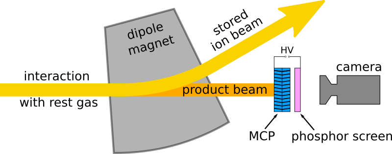
The neutral imaging setup at CRYRING is used for beam imaging of singly-charged ion beams. It utilizes the intrinsic beam loss process of charge exchange with residual gas that projects a two-dimensional beam profile onto a multi-channel plate detector with a phosphor screen. This provides a much needed diagnostic tool for setting up optimal electron cooling and for experiments using singly charged ions. Below you can see an example of electron cooling of an Mg+ beam observed with the setup. The beam is injected, moves slightly to the outside of the ring during acceleration and is cooled which leads to a reduction in the diameter.
Measurement Principle
The existing ionization profile monitors (IPMs) at CRYRING cannot be used for singly-charged ions without destroying the beam. Therefore a non-destructive system was needed and we decided to use the process of charge exchange which in the case of singly-charged ions leads to a neutral product beam that passes through the dipole bending magnet behind each straight section without being affected. We can then image this product beam and get an accurate measurementof the beam profile in the preceding straight section. This is especially useful when trying to establish electron cooling. The detector setup is positioned inside the vacuum behind the dipole magnet in section YR05. It consists of two MCP plates in a chevron configuration and a phosphor screen. A camera is positioned outside the vacuum and captures the fluorescence on the phosphor screen that results from the impact of a neutral particle on the MCP and the subsequent electron cascade.
Pin assignment
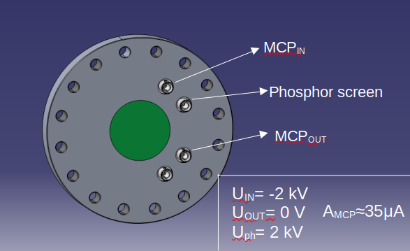
The assignment of the pins on the MCP flange, as well as the voltage applied on each pin and the corresponding electrical current are shown in the sketch below.
| I | Attachment | Action | Size | Date | Who | Comment |
|---|---|---|---|---|---|---|
| |
ElectronCooling.png | manage | 1 MB | 2022-03-09 - 17:25 | EstherBabetteMenz | Observation of electron cooling on the neutral imagin setup |
| |
MCP.png | manage | 51 K | 2023-10-26 - 15:02 | ElenaOanaHanu | |
| |
neutralimaging_setup.png | manage | 46 K | 2022-03-09 - 17:20 | EstherBabetteMenz | Schematic of the neutral imaging setup |
Edit | Attach | Print version | History: r5 < r4 < r3 < r2 | Backlinks | View wiki text | Edit wiki text | More topic actions
Topic revision: r5 - 2023-10-26, ElenaOanaHanu
 Copyright © by the contributing authors. All material on this collaboration platform is the property of the contributing authors.
 Ideas, requests, problems regarding GSI Wiki? Send feedback | Legal notice | Privacy Policy (german)


